The EYE is a vital visual organ. It is the organ that makes it possible for us to SEE and appreciate the beauty of the world. Without the eyes, we will be in the ‘dark’ and blind to everything around us.
The EYE is a complex organ. It rests safely in a socket and referred to as an ‘eyeball’. This “ball” is made of optical parts, tissues, tiny muscles, blood vessels etc. These parts are delicate and light-sensitive, and work together to send visual stimuli ( message ) to the brain through a link nerve called the Optic Nerve. This process which makes it possible for us to SEE, takes place in split seconds. To understand this process -( how the eyes function ), it will be necessary to know the basic parts of the eyes.
PARTS OF THE EYE
The eye is made up of external and internal parts.
The External Parts
These are the parts of the eyes that are visible to you in the mirror. I love to call it “the eye in the mirror”. These consists of
Conjunctival — an external part of the eye. It is a thin clear and transparent layer covering the white part of the eye called the sclera.
Sclera —- the white part of the eye. It may appear white in its natural healthy state. However it has tiny blood vessels running through it.
cornea. —-is literally the most external part of the eye. It is a clear and transparent tissue through which the pupil and iris are visibly seen. Based on its transparency and clear visibility of the iris, most people have wrongly referred to it as the ‘black part’ of the eye.
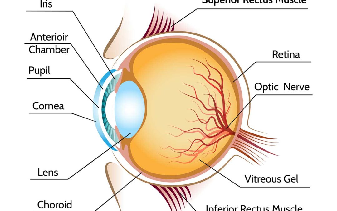
The Iris — is referred to as ‘the black part” of the eye’. However, depending on race and genetic make up of people, the iris could be brown, gray, blue or green. The iris functions like the shutter of a camera. Its reaction to light alters its shape and causes an increase or decrease ( dilate or constrict ) in the size of the pupil.
The pupil — is an opening through which light passes to the back of the eye. It is visibly located in the centre of the iris and naturally darker than the iris. It functions like the aperture of a camera. Its size changes according to the amount or intensity of light exposed to it. It controls the amount of light that goes into the eye. Just like a check valve, it ensures that not too little or too much light gets to the back of the eye.
The Internal parts consist of
The lens – a small-size clear lens that has the ability to alter its shape depending on the distance of your focus. The shape alteration makes it possible for light rays to be focused on the retina for a clear distance or near vision as required.
The segments – This is divided into two; the front ( anterior ) segment which is filled with aqueous fluid and the back ( posterior ) segment which is filled with a jelly-like fluid called the vitreous. The vitreous helps maintain the shape of the eyeball and refract light rays that pass through it to the retina.
The retina – The retina is the back of the eye and the part of the eye where light rays comes into focus. It is a layer of tissue with many micro internal parts; blood vessel, macular, nerve endings, photo receptors. etc. and connected to the brain through a delicate nerve called the optic nerve. Through this nerve, the visual stimuli and impulse from the retina are relayed to the brain.
It should be noted that the brain interprets what we ‘see’.
HOW THE EYES WORK
Everyone knows the function of the eyes but not everyone knows how it functions. How does the eye carry out its visual activity? You may have wondered. Well, it all starts from the light in your environment.
Light is reflected from the objects in your surrounding and passes through the cornea to the first segment ( filled with aqueous fluid). And through the pupil, it gets to the Lens and then to the second segment which is filled with a jelly-like fluid called the vitreous. The vitreous fluid is what maintains the shape of the eyeball. From the vitreous, the light hits the retina where the photocells (photoreceptors) convert it into electric-visual stimuli.
This stimuli travels through the optic nerve to the brain for processing. You may relate this to ‘computer processing’. The part of the brain that does the processing is the visual cortex which converts the visual stimuli into an image and interprets the image. The interpretation of the image by the brain is what makes it possible for us to identify or SEE details of the things in our view such as shape, size, colour, clarity, contrast, proximity, movement etc due to the link or connection between the eyes and the brain.
Written By – Austin Madu, OD (Optometrist )
Recent Comments