Your Eyes – What You Need To Know

Your Eyes – What You Need To Know

  The EYE is a vital visual organ. It is the organ that makes it possible for us to SEE and appreciate the beauty of the world. Without the eyes, we will be in the ‘dark’ and blind to everything around us. 

The EYE is a complex organ. It rests safely in a socket and referred to as an ‘eyeball’. This “ball” is made of optical parts, tissues, tiny muscles, blood vessels etc. These parts are delicate and light-sensitive, and work together to send visual stimuli ( message ) to the brain through a link nerve called the Optic Nerve. This process which makes it possible for us to SEE, takes place in split seconds. To understand this process -( how the eyes function ), it will be necessary to know the basic parts of the eyes.


                          PARTS OF THE EYE

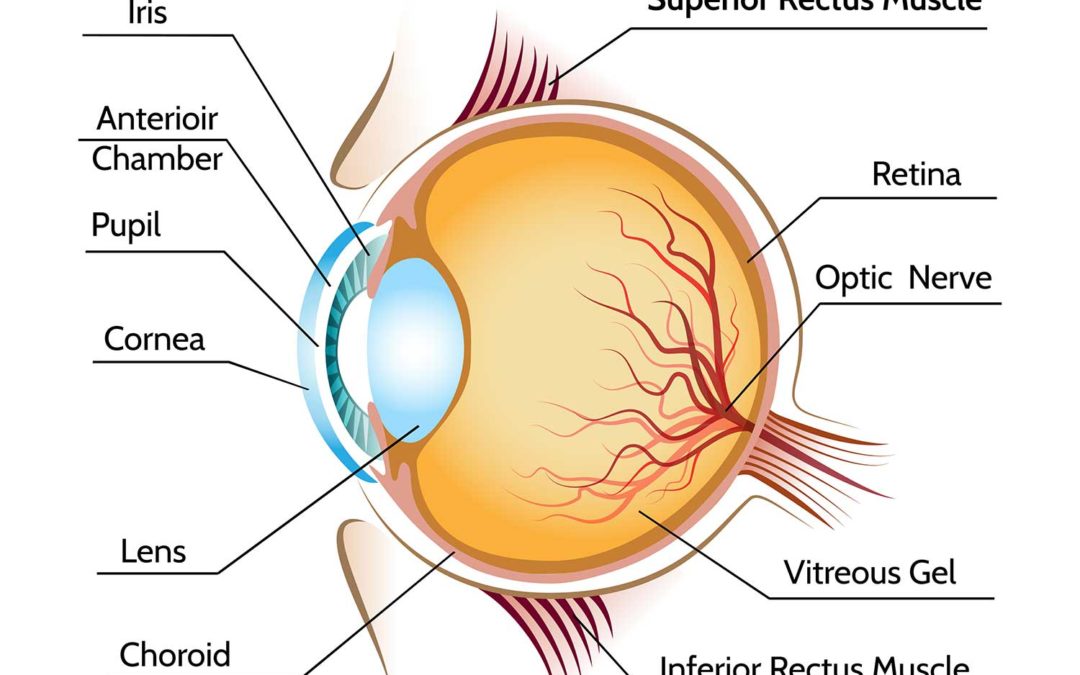
 The eye is made up of  external and  internal parts. 

The External Parts

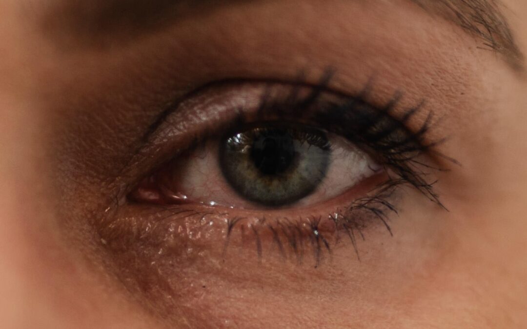
These are the parts of the eyes that are visible to you in the mirror. I love to call it “the eye in the mirror”. These consists of

Conjunctival  — an external part of the eye. It is a thin clear and transparent layer covering the white part of the eye called the sclera.

Sclera —- the white part of the eye. It may appear white in its natural healthy state. However it has tiny blood vessels running through it.

cornea. —-is literally the most external part of the eye. It is a clear and transparent tissue through which the pupil and iris are visibly seen. Based on its transparency and clear visibility of the iris, most people have wrongly referred to it as the ‘black part’ of the eye.

The Iris — is referred to as ‘the black part” of the eye’. However, depending on race and genetic make up of people, the iris could be brown, gray, blue or green. The iris functions like the shutter of a camera. Its reaction to light alters its shape and causes an increase or decrease ( dilate or constrict ) in the size of the pupil. 

The pupil — is an opening through which light passes to the back of the eye. It is visibly located in the centre of the iris and naturally darker than the iris.  It functions like the aperture of a camera. Its size changes according to the amount or intensity of light exposed to it. It controls the amount of light that goes into the eye. Just like a check valve, it ensures that not too little or too much light gets to the back of the eye.

The Internal parts consist of

The lens –  a small-size clear lens that has the ability to alter its shape depending on the distance of your focus. The shape alteration makes it possible for light rays to be focused  on the retina for a clear distance or near vision as required.    

The segments – This is divided into two; the front ( anterior ) segment which is filled with aqueous fluid and the back ( posterior ) segment which is filled with a jelly-like fluid called the vitreous. The vitreous helps maintain the shape of the eyeball and refract light rays that pass through it to the retina.

The retina – The retina is the back of the eye and the part of the eye where light rays comes into focus. It is a layer of tissue with many micro internal parts; blood vessel, macular, nerve endings, photo receptors. etc. and connected to the brain through a delicate nerve called the optic nerve. Through this nerve, the visual stimuli and impulse from the retina are relayed to the brain.

It should be noted that the brain interprets what we ‘see’. 

            HOW THE EYES WORK 

Everyone knows the function of the eyes but not everyone knows how it functions. How does the eye carry out its visual activity? You may have wondered. Well, it all starts from the light in your environment.

Light is reflected from the objects in your surrounding and passes through the 
cornea to the first segment ( filled with aqueous fluid). And through the pupil, it gets to the Lens and then to the second segment which is filled with a jelly-like fluid called the vitreous. The vitreous fluid is what maintains the shape of the eyeball. From the vitreous, the light hits the retina where the photocells (photoreceptors) convert it into electric-visual stimuli.

This stimuli travels through the optic nerve to the brain for processing.  You may relate this to ‘computer processing’. The part of the brain that does the processing is the visual cortex which converts the visual stimuli into an image and interprets the image. The interpretation of the image by the brain is what makes it possible for us to identify or SEE details of the things in our view such as shape, size, colour, clarity, contrast, proximity, movement etc due to the link or connection between the eyes and the brain. 

      Written By – Austin Madu, OD                                                (Optometrist )

Dry Eyes – What You Need To Know

Dry Eyes – What You Need To Know

Dry Eyes is an eye condition that affects millions of people. This condition affects more women than men and more of the older than the younger people. Chronic Dry Eyes ( CDE) is simply referred to as Dry-Eyes (DE ) and has a lot to do with tears production of the eyes. However, It is important for you to know that tears is NOT water. Tears contain, in addition to water, lubricants, nutrients and electrolytes. This content is what gives the tear-film its stability, lubricating, moisturising and cleansing functions that it provides to the surface of the eyes each time we blink.

Symptoms of Dry Eyes 

The symptoms of dry eyes may be temporary and occasional for some patients, but severe and worrisome for others. The common symptoms of dry eyes are dryness of the eyes, itchy-burning sensation, Gritty sensation (your eyes feel like you have sand particles in them ), increase in light sensitivity, off and on blurry vision etc. As surprising as it may sound, some patients with DE may experience ‘excessive tearing’. Yes , ‘Tearing’…..Now you may wonder why a patient with dry eyes would have watery eyes. The answer lies in the CAUSE of the Dry Eye condition. 

Causes Of Dry Eyes

Dry Eyes is most commonly caused by insufficient production of tears. This is a case where the eyes are not able to produce enough tears to lubricate the eyes. In this instance, the eyes produce healthy but very SCANTY tears – “Quality with little QUANTITY. “ 

Dry Eyes can also be caused by deficiency of tear content. In this instance, the tears lack its natural content or composition. The eyes produce sufficient but unhealthy tears – “Quantity with NO QUALITY”. As a result of this deficiency, the tears become UNSTABLE and unable to bond to the surface of the eyes when we blink and therefore washes off like water to the lower portion of the eyes, leaving the eyes with no lubrication and causing dryness. This triggers more ‘watery’ tears production and the cycle repeats. In this case, patients with Dry Eyes will experience ‘watery’ eyes with some sort of discomfort.

Health Conditions That Can Result To Dry eyes  

 – Hormonal changes; This is common with women due to pregnancy, menopause etc

– Poor Health condition; diabetes, high level of cholesterol, thyroid, cancer etc    

 – Ageing process of the body; This leads to degeneration of most parts of our body including the eyes.         

– Side effects and prolonged use of certain medications.

-Eye imflammation; This may have affected the tear gland ( the gland responsible for tears production )     

-Certain medical procedures;  For example Chemotherapy radiation treatment for cancer patients.   

Environmental condition; prolonged, constant and unprotected exposure to harsh environmental condition such as heat, direct sunlight, toxic chemicals or fumes.

Some medical  authorities have claimed that the use of computer causes Dry Eyes. This is not true. The fact is that even though prolonged use and constant focus on the computer or any electronic device without frequent blinking could temporarily make the eyes dry. This effect does not cause or trigger Chronic Dry Eyes ( kerato-Conjunctivities Sica ) . However, it could worsen the symptoms of Dry Eyes if it is not managed with an eye drop.

Treatment

Chronic dry eye is untreatable. But as frustrating as this may seem, the good news is that it is manageable. There are a number of OTC eye drops for dry eyes. Your eye doctor should recommend one for you if you have DE. However the OTC drops that I recommend for most of my patients are quite effective and you can get them in your local pharmacy stores.

Written By;     

Austin Madu,  OD   

(Optometrist )

Why Your Near Vision Gets Blurry As You age

Why Your Near Vision Gets Blurry As You age


As people advance in age, numerous changes take place in the eyes. One common effect of these changes is blurry near-vision. This age-related vision condition is known as presbyopia. It starts slowly, progresses gradually and becomes noticeable from an early to mid 40s and continues to the 60s. It is caused by the loss of the eyes ability to focus on near objects especially objects within 50 cm distance-range from the eyes. The closer an object or fine prints are from the eyes, the more blurry and strenuous it becomes for the eyes to see. This is the reason people with this condition ( presbyopic patients ) hold books or small items with their arm stretched forward in order to see clearly.


The Changes In The Eyes That Cause Presbyopia

The two parts of the eyes responsible for this condition are the lens ( which is naturally flexible ) and the ciliary muscles to which the lens is attached. Before late 30s, the ciliary muscles are effortlessly responsive to constant change of focus between far and near objects/prints. Its responsiveness alters, in split seconds, the shape of the lens which is naturally flexible.

However, as we get older, the ciliary muscles and lens, like most part of our body start to get weak. In other words, the ciliary muscles start to lose its responsiveness and the lens starts to lose its flexibility which makes it difficult for image of a tiny object or fine prints at close range to be focused on the retina and therefore hard for a presbyope to read fine prints or thread the needle. This whole process of constant change of focus from far to near or near to far is known as accommodation.

In a nutshell, as we get older, the eyes tend to lose accommodation which results to presbyopia and which as I earlier said, is age-related and nothing to worry about.

If you have presbyopia I advise that you visit an optometrist for eye examination to get prescription glasses. This may be progressive or bi-focal lens.                   

Written By;

Austin Madu, OD                           

Optometrist

Is Carrot Good For Your Eyes

Is Carrot Good For Your Eyes

.Is there  any relationship between carrot and the eyes?

Why do most people associate carrot with good eye-sight or believe that it will improve their vision.? 

These are some of the questions, you must have asked or may want some answers to.

Firstly, let’s take a look at the content and key ingredient of carrot- carotenoid.

Carotenoid
Carotenoid is the key ingredient of carrot. It is a fat soluble antioxidant plant compound found both in carrots and many other vegetables that you consume on daily basis. 

When foods that are rich in carotenoid are consumed, carotenoid is converted into two groups of compounds.

1) The carotene group ( common among this group is a type of compound called beta-carotene which is converted in the human body to Vitamin -A )

2) The xanthophyll group ( this is converted to Lutein and zeaxanthine. These compounds are predominantly found in the retina (back of the eye ).

Vitamin-A                                   

Vitamin A is essential in keeping the retina and photoreceptors healthy. The photoreceptors are photocells in the retina that are responsible for day light and night vision. Deficiency of Vitamin A can affect the functions of these photocells and could lead to poor night vision. Prolonged deficiency of  vitamin -A in the body could result to a condition called xerophthalmia and night blindness. This condition is often common in highly impoverished communities where there is scarcity of food or where people eat very poor diet. 

Lutein and xeaxanthine 

These compounds have antioxidant functions which help slow down the rate of degenerative changes that take place in the eyes resulting from aging process and the presence of free radicals in the body. These changes often lead to eye conditions such as age-related macular degeneration (AMD ) and cataract.

Like all vegetables that are rich in vitamins, carrot is good for your eyes and for other parts of your body too. But before you hurry down the road to the vegetable shop to buy some carrots, you need to know that the amount of carotenoid the eyes need to function properly is not only found in carrot. It can also be found in other foods that you consume. Some of these are Tomatoes, Water melon, Kale, Spinach, Sweet potatoes, Tangerine, Pumpkin, and many yellow-coloured fruits and vegetables.

This implies that carrots are not one-of-a-kind or vision-saver vegetables.

I have not come across any clinical evidence that showed that consumption of carrots alone prevented the development of cataract, macular degeneration or improved a person’s eyesight and making the use of glasses unnecessary.

When it comes to healthy eyes and good eyesight, factors such as – aging, genetics, nutrition, over-all health and medical conditions etc matters the most.

Written By;

Austin Madu, OD 

( Optometrist )


Contact Lens Guidelines, Dos And Don’ts

Contact Lens Guidelines, Dos And Don’ts

 For a safe and comfortable contact lens, an important factor that every contact lens wearer should take note of is hygiene.

However to ensure proper contact lens hygiene, there are guidelines that a contact lens wearer must follow.


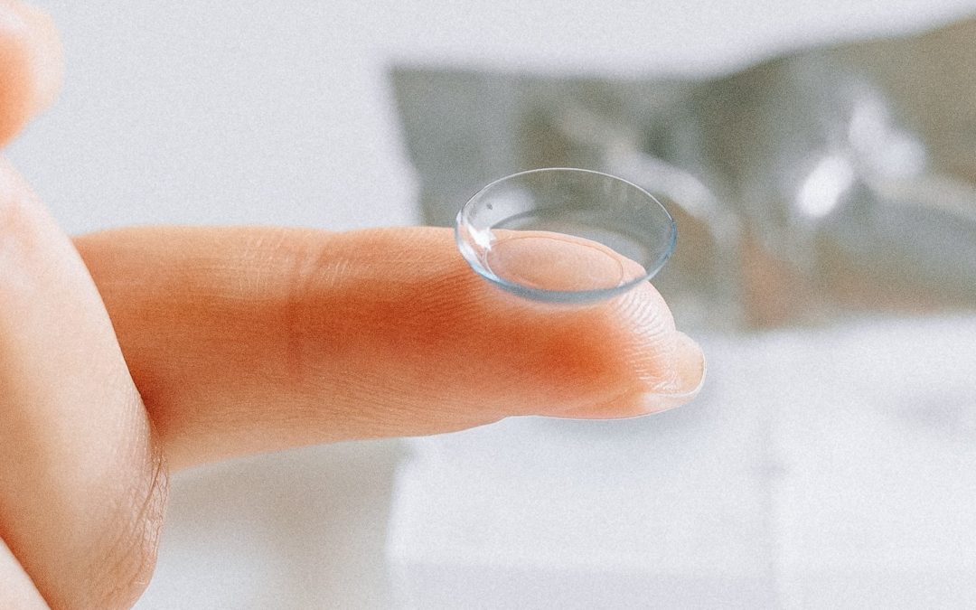
1) Before taking out your contact lens (CL) from its case, wash your hands properly with soap and water and dry-clean them with a clean hand towel. (use this towel only for CL)


2)  To avoid mixing them up, make it a rule to always apply the right-eye CL on the right eye first and then the Left-eye CL on the left eye. If you choose the opposite, its not a problem. Make sure that you are consistent with it. 


3) When you apply CL on the cornea (eye), blink several times. This helps center the lens and remove trapped air bubbles. Take a minute or two to observe how you feel. If you feel comfortable, you are ready to “go”. Now empty the lens case and cover it . Do not leave it exposed to avoid contamination. However, if you feel uncomfortable after applying the lens, remove it immediately and inspect it to make sure that

(i) There is no particle on it. If there is, it should be rinsed out with CL solution.

(ii) The lens is not turned inside-out. If it is, carefully invert it

(iii) There is no tear. If there is, it should be discarded.


4) If you experience dryness of the eyes when wearing your CL, apply re-wetting solution. Avoid stepping into dusty, smoky, gaseous or hazardous  environment as much as possible. These environments could cause eye irritation and in some cases serious eye infection.  


5) At the end of the day (before going to bed) remove the lens from your eyes.  Remember to wash and dry your hands before you remove the CL from your eyes. To avoid mixing them up, remove Right CL first and rest it gently in the ‘right-side’ of the CL case. Do the same for the left eye. Now fill the case with the CL solution- enough to completely cover the lenses.

In addition to these basic guidelines, it is crucial to know and adhere to the do’s and dont’s of contact lens wear.


Do’s and Dont’s of Contact Lens wear

— A first-time contact lens ( CL ) wearer should not wear it for more than 2-3hrs the first day. A gradual wearing ‘build-up’ time as advised by the optometrist should be followed.

–Regardless of the type of CL you have, I DO NOT advise that you go to bed wearing CL. This is because the cornea (your eyes ) are better oxygenated without CL on them while you sleep. And also better rejuvenated the next morning. This also makes the cornea less prone to CL-induced-infection.

–Carry your CL solution and case with you because you may need it during the day.

–Never wash your CL with water. This can cause serious eye infection by an organism called ‘acantamoeba’.

–Never leave your CL in its case without CL solution in it. This could deform the lens and render it useless.

–Do not use your CL and case more than the recommended disposable time. CL case should be changed at least every three months.

–Women should take extra care when applying make-ups, eye liner etc.

–Whatever be the case if you experience redness, pain, irritation, swelling, itching etc  please remove the CL immediately and see your optometrist/ophthalmologist


Please always follow the advise and recommendations of your eye care professional.                                        

 Written By: 

Austin Madu, OD                                             

 ( Optometrist )